by Karen O'Hanlon Cohrt | Mar 22, 2026 | Disease Models, Trends
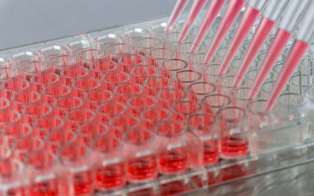
It is no secret that cell-based models are the backbone of modern drug discovery. For decades, researchers have used animal-based models including cells and whole animals to investigate mechanisms of disease, screen compounds, and support preclinical research. Over...
by Karen O'Hanlon Cohrt | Jan 6, 2026 | Cell Culture Techniques, Disease Models
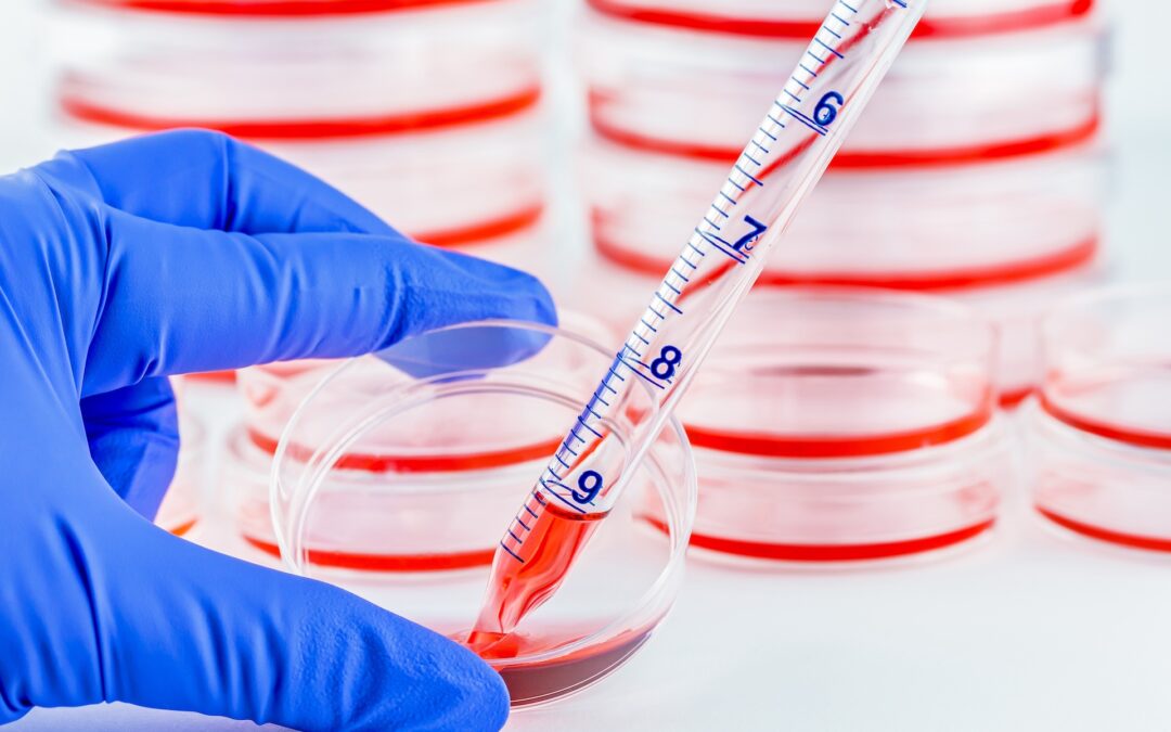
What is the difference between ECM-based and ECM-free systems in 2D and 3D cell culture? When are ECM-derived cues required for tissue organization and differentiation in 3D models? How do researchers choose between ECM-based and ECM-free culture formats for their...
by Karen O'Hanlon Cohrt | Nov 6, 2025 | Cell Culture Techniques, Disease Models
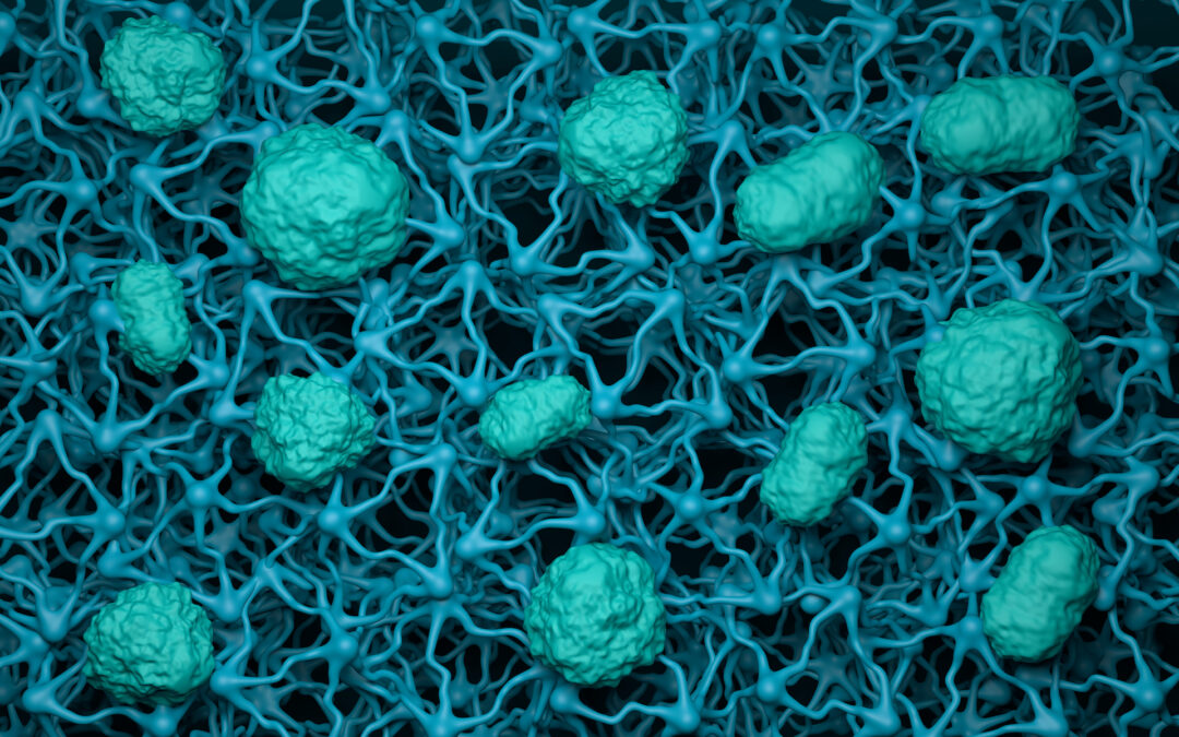
How do Transwell assays help model biological barriers in the lab? What does TEER tell us about barrier integrity and tight-junction strength? How can these assays advance our understanding of drug transport and disease? Biological barriers are critical for...
by Karen O'Hanlon Cohrt | Oct 6, 2025 | Disease Models, Trends
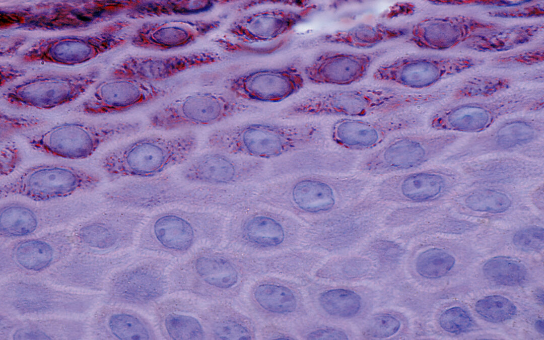
In a previous article we introduced keratinocytes and looked at their biological functions and subtypes. Here, we explore some of the main reasons researchers study keratinocytes and the various approaches used. We focus on 2D assay formats, outlining their advantages...

by Olwen Reina | Sep 13, 2016 | Disease Models
What are keratinocytes? Take a look at your hands, your face and your toes. Most of what you’re seeing are your keratinocytes. They make up over 90% of the cells of the epidermis, the outermost layer of the skin. The skin on your neck and the soles of your feet,...

by Allison Kennedy | Jun 26, 2023 | Disease Models
Liver sinusoidal endothelial cells (LSECs) are highly specialized liver endothelial cells that form a physical barrier between the blood and hepatocytes. They are the most abundant non-parenchymal hepatic cell population. LSECs play an important role in physiological,...