What are keratinocytes?
Take a look at your hands, your face and your toes. Most of what you’re seeing are your keratinocytes. They make up over 90% of the cells of the epidermis, the outermost layer of the skin. The skin on your neck and the soles of your feet, the underside of your arm and your knees is very different. This difference is mainly in toughness and is caused by the amount of keratin proteins produced by the differentiated keratinocytes in that part of your skin. Keratin is an intermediate filament protein produced by keratinocytes.
The role of keratinocytes in the skin
The main purpose of these keratin-producing cells is to preserve against microbial, viral, fungal and parasitic invasion; to protect against UV radiation; and to minimize heat, solute and water loss. They are used to research a number of phenomena of the skin including epidermal acidification, DNA degradation, fatty acid metabolism and transport, local immune responses, cell regeneration, stem cell differentiation and tumor formation.
The lifecycle of the keratinocyte
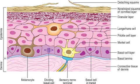
To recap, the skin is divided up into three layers: the epidermis, the outermost layer of skin; the dermis, directly under the epidermis; and a subcutaneous or fat layer, under the dermis. The epidermis can be further divided into sublayers:
- the basal lamina (the innermost layer)
- the basal cell layer
- the prickle cell layer
- the granular cell layer
- the keratinized squames (the outermost layer)
Before we look at the types of keratinocytes, we’ll first look at an overview of the lifecycle of a keratinocyte. A keratinocyte can have two fates:
- to be a dividing cell in the basal layer, or…
- to begin differentiating and migrating through the layers of the skin.
We’ll look at both processes here.
In the basal layer of the skin, the innermost stratum, a basal keratinocyte has just divided by mitosis to form a new basal keratinocyte. This new cell starts to divide itself and produces many more keratinocytes. Some of these cells will stay with their parent and continue to replenish the population of basal keratinocytes. These cells are known as stem cells. However, other cells will start a process of differentiation.
Over time, the these differentiating cell are pushed upwards as the next generation of cells forms underneath them. Eventually, they are pushed into the next layer of the skin to become prickle cells. As more and more cells are made in the basal layer, the newly formed prickle cells continue getting pushed upwards and eventually they reach the granular layer. Here, the cells undergo a semi-apoptotic series of events in which their cell organelles and nucleus are degraded overtime. Once they’ve been pushed into the highly keratinized squamous layer and become squames. These cells are very flat and eventually they flake off as dead skin cells. With each stage, the cells produce a different profile of keratin proteins in a process known as terminal cell differentiation. This process is illustrated in this video on keratinocytes.
Depending on the region of the body, this lifecycle can take about a month. Over the course of a lifetime, the skin in renewed approximately a thousand times. Not all cells on the basal cell layer will end up as squames, since some are needed to maintain the cell population. To ensure there is an aquate number of cells both dividing and differentiating, the balance between the keratinocyte stem cell population and the cells destined to become terminally differentiated must be maintained. Usually, so long as an approximately equal number of cells are being created for both populations, this balance is maintained. As you can imagine, this involves an intricate balancing act with a lot of players involved to keep the peace!
The types of keratinocytes
Keratinocytes change in appearance from one layer of the skin to the next. They start in the basal cell layer and migrate upwards. Those in the lowest stratum, or layer, of the skin are called basal cells. These are usually the only ones that divide.
Above these are several layer of larger prickle or spinous cells. Prickle cells are held tightly to one another by intercellular attachment points called desmosomes. Each desmosome is composed of membrane proteins that allow the cells to link together. These membrane proteins are in turn bound by anchoring proteins that form a disc-shaped plaque on the inner surface of the membrane. The anchoring proteins are bound by keratin filaments. These desmosomes appear under light microscopy as spiky cell membrane projections giving the cells a prickly appearance, a little bit like a thistle plant, hence the name prickle cells.
Above the prickle cells are the granular cells. This layer forms the waterproof barrier characteristic of the skin. This protective barrier is the boundary layer that separated the inner, metabolically active strata and the outer highly-keratinized, dead layers of the skin.
Above the granular cells are the squames. These extremely flattened cells are highly keratinized meaning they’re extremely densely packed with keratin protein. Both the squames and the outermost layer of the granular cells just below the squames are armored with 12nm-thick, cross-linked layers of protein.
In our next article, we’ll look at the role of keratinocytes in healing and examine how the balance between the types of keratinocytes is maintained. We’ll also look at how keratinocytes work together with melanocytes as well as the role they play in disease. Lastly, we’ll examine how keratinocytes are being used commercially in soft tissue regeneration.
If you’d like us to cover any other topics, send us an email or leave a comment below.