Organoids are cell-derived, 3D in vitro models that are cultured to recapitulate structural and functional aspects of the in vivo tissue they are intended to represent. Organoids are not new to research labs, although there has been much hype about them in recent years, given their vast and largely untapped potential to reveal novel insights about biological processes and disease mechanisms, as well as serve as models for drug discovery and development, including drug target identification and validation as well as pre-clinical toxicity and efficacy studies.
In previous articles, we looked at the history of organoids, and outlined the typical workflow for their development from pluripotent stem cells. We also looked at their uses in research and development, with a particular focus on cancer organoids. More recently, we wrote about human liver organoids as an emerging model for non-alcoholic fatty liver disease.
In this article, we take a look at some of the major considerations for organoid culture, and summarize the quality control methods used to evaluate organoid integrity: i.e. how well they actually represent the structure and function of the organ or tissue they are intended to mimic.
What are the considerations for organoid culture?
Among the main considerations for organoid culture are soluble factors (primarily growth factors or small-molecule drugs that modulate various signaling pathways), physical and integrating cues, cell source and matrix. As outlined in a previous article about providing the right cues for organoid development, the exact combination of soluble factors and cues necessary are tissue-specific, and will not be discussed in detail here. Instead, we will cover cell source and matrix as two of the critical factors governing successful organoid culture.
Cell source is critical because the starting cellular population used to culture any organoid will dictate the resulting variability and heterogeneity in the organoid, as well as the function of the tissue or organ the organoid is intended to mimic. The term matrix within organoid technology refers to the addition of extracellular matrix (ECM), which is often a gel-based material that offers structural support as well as some biochemical cues to the growing organoid.
Getting the desired cells out of the source tissue
For tissue-derived organoids or cancer organoids, tissue-resident stem, progenitor, or differentiated cells or tumor cells are isolated using highly optimized dissociation methods, which often include the addition of tissue-specific enzyme cocktails to degrade the natural extracellular matrix, as well as EDTA to chelate calcium and disrupt cell-cell adhesion and tissue integrity. In some cases, DNAse addition may be necessary to eliminate excessive DNA that escapes from ruptured and necrotic cells. Additional enzymatic steps may also be incorporated to yield single-cell suspensions if desired. For tumor-derived organoids, cell clusters are preferable to single cells.
It is important to note that enzymatic isolation methods may impact the condition of the obtained cells especially when longer enzymatic incubation periods are required. Mechanical dissociation methods, which include a combination of aspiration, vortexing, scraping, or tissue pressing, offer a faster and cheaper alternative to enzymatic methods, albeit with the risk of inconsistent cell yields and viabilities. In some cases, selective culture media may be used to enrich certain cell types.
In practice, a combination of enzymatic and mechanical methods are often used to dissociate tissues for organoid culture, and the exact method chosen should consider the sensitivity of the cell type being isolated and how much ECM it naturally produces; cells that secrete excessive ECM will likely be more difficult to remove from cell culture plates.
Tissue-specific adult stem-cell derived organoids (whether from healthy or diseased patients) are isolated from their source via similar dissociation methods as described above, and then embedded into a 3D matrix designed to mimic the relevant stem cell niche, i.e., the in vivo microenvironment in which stem cells reside and receive stimuli that govern their fate.
Once the dissociation process is complete, regardless of cell source, the isolated cells are sorted by analyzing for tissue-specific cell markers or specific physical features so that only the desired cell type(s) are used to seed the organoid. Methods used here typically include FACS or magnetic cell separation using specific antibodies or ligands to isolate cells of interest.
iPSCs are reprogrammed and cultured as 2D monolayers. Currently, most labs culture iPSCs without feeder cells (which are usually mouse embryonic fibroblasts or MEFs) and use different coating materials and methods to support their growth and maintenance. Human iPSC-derived organoids are cultured as a differentiated unit of multi-cell-types in 3D. The iPSC derived organoids may not require a physical 3D matrix to support them
Choosing the right matrix
The main functions of the matrix are to provide structural support such as porosity and stiffness, as well as signaling cues that promote cell migration, cell behavior and polarization in organoid structures. According to Zhao et al. (2022), the ideal matrix should be stress-reducing and offer highly dynamic biochemical and biophysical properties to allow for or modulate necessary changes in organoid structure during culture.
Commonly used matrices include the biologically-derived Matrigel as well as synthetic hydrogels or recombinant human collagen. Matrigel is a soluble extract of basement membrane proteins secreted by Engelbreth-Holm-Swarm mouse sarcoma cells that resembles the complex extracellular environment of most tissue types. Matrigel naturally contains laminin, collagen IV, enactin, perlecan and growth factors, and is probably the most widely used matrix material in organoid culture. Matrigel is commercially available in various formats, including a version that is optimized for organoid culture. Organoid-grade Matrigel products are typically verified to support murine and human organoid culture, are tested for their ability to form and maintain stable 3D droplets on multi-well plates, and are measured for matrix stiffness.
Although Matrigel has long been seen as the golden standard matrix material within organoid biology, it is an animal-based and heterogeneous substance with a variable composition that offers limited control over biochemical and biophysical spatio-temporal cues that are essential to streamline organoid culture. These limitations have sparked efforts in the field to develop reproducible alternatives including those mentioned above, as well as fibrins that may be supplemented with laminin-111 and factor-XIII to more accurately mimic natural ECM (Broguiere et al., 2018).
Recognizing those shortcomings of Matrigel, chemical companies have developed novel synthetic hydrogels based on polymers such as polyethylene glycol, nanocellulose, alginate, hyaluronic acid, and others (reviewed in Poudel et al., 2022). One notable attraction of a fully synthetic ECM is the possibility to manipulate and fine-tune matrix properties by adding or omitting certain structural components or signaling cues from the matrix ‘recipe’, or by controlling the degradation of the matrix which in turn influences the cell behavior. For instance, Kloxin and co-workers demonstrated that photodegraded channels within a photodegradable PEG-based hydrogel containing encapsulated permitted cell migration (Kloxin et al., 2009).
Despite progress in alternative matrix technologies, Matrigel is still considered the universal ECM product for organoid growth and organoid culture overall remains to be more efficient when Matrigel is used, highlighting the need for further advances in this field.
So the organoids are growing, but how ‘good’ are they?
Organoid culture is a multi-step process that involves initial aggregation (if single cells are used), proliferation, migration and differentiation. Before the organoids can be used experimentally, e.g., to address research questions or test new drugs, it is imperative to confirm that 1) they actually contain the desired cell type(s), and 2) that they exhibit the gene expression signatures and recapitulate the features and functions of the organ they are intended to mimic, and to what extent they do this.
In a nutshell, organoid characterization is a major challenge for the field, and no consensus exists with respect to (universal) biomarkers, which assays should be used, and what criteria must be satisfied before an organoid can be considered to mimic the target organ in vivo. Much of the trouble with characterization stems from massive gaps in our understanding of the target organs themselves. How can one mimic something if they don’t know exactly what it looks like/how and why it functions the way it does? Organoid biology is evolving as new assay and imaging technologies emerge, but for now we will take a general and simplified look at the approaches to organoid characterization in use today.
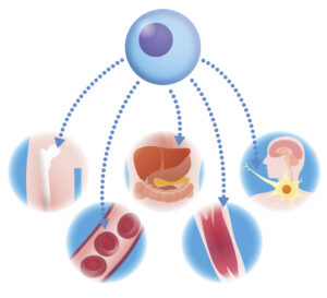
Illustration: Human iPSC-derived organoids.
Does it look like the target organ in vivo?
These quality control steps can be tackled in various ways, and usually involves a combination of low-throughput gene expression validation analysis (e.g., real-time RT-PCR) and high-throughput transcriptomics to analyze gene expression, which together answer the question: does the organoid exhibit the expected gene expression signature, i.e., are key transcription factors and cell-specific markers expressed? Single-cell RNA sequencing can be useful in revealing the extent of cell differentiation within an organoid.
Immunoassays including western blotting and immunoprecipitation shed light on protein abundance, protein-protein interactions and post-translational modifications – do these mimic the patterns of the intended organ? In addition, immuno-qPCR, which combines the signal amplification power of real-time PCR with the robustness and specificity of ELISA-based immunoassays allows for the detection of organ-specific biomarkers with dramatically increased sensitivity compared to conventional ELISA.
Immunofluorescence and immunohistology further supplement organoid characterization by allowing side-by-side morphological and staining comparisons of organoid vs. organ-derived sections. Here, it is critical to confirm that the organoid is structurally similar to the target organ, e.g., pancreas-derived organoids should possess an islet-like globular structure and intracellular hormone vesicles, while crypt-like structures should be readily visible in intestinal organoids. However, since structure and shape are difficult to define, especially for organs that are less well understood in detail, morphology is difficult to reproduce at scale and this is a significant challenge for those in the field.
Does it perform the expected functions?
No organoid will fully recapitulate the gene expression signatures, morphology or functionality of an in vivo organ, but characterizing organoid functionality in comparison to freshly prepared tissue is essential for quality control prior to downstream experiments.
The exact criteria used to assess organoid function will be organ-specific, but typically include parameters such as: cell maturation, vascular development, formation of neuronal networks (e.g. in brain organoids), secretion of expected cytokines (e.g. in organoids that incorporate immune cells) and hormones (e.g. endocrine organoids), acid secretion (e.g. stomach organoids), mucus secretion (e.g. intestinal organoids), calcium signaling capabilities (e.g. muscular organoids such as the heart, skeletal muscle and retina), and enzymatic activity (often used to determine liver-like functionality).
Assays used to determine organoid functionality include real-time PCR, high-throughput RNA sequencing, immunoassays, microscopy in combination with staining, as well as various biochemical assays to measure enzymatic and signaling activities.
A comprehensive overview of the methods used to determine organoid structure and function can be found in a recent review by Zhao et al. (2022).
As complex 3D mixtures of cell types, organoids are often considered to be superior to all other culture methods, but they might not alway be the best solution in practice. Before costly and time-consuming attempts are initiated to set up an organoid culture, the experimental goals should be scrutinized. For instance, because organoids are 3D structures, they move and rotate, which poses challenges for long-term live-imaging experiments. In addition, problems with organoid reproducibility continue to hamper scaled-up applications. In such cases, a simpler 2D or 3D co-culture that is established using microfluidics, Transwells, or other similar devices may be useful (see Hofer and Lutolf (2021) for a review on this topic).
That was it for now! Stay tuned for a future article where we will overview some of the recent advances related to organoid applications in research, drug discovery and development.
References:
- Zhao Z, Chen X, Dowbaj AM, Sljukic A, Bratlie K, Lin L, Fong ELS, Balachander GM, Chen Z, Soragni A, Huch M, Zeng YA, Wang Q, Yu H. Organoids. Nat Rev Methods Primers. 2022;2:94. doi: 10.1038/s43586-022-00174-y.
- Broguiere N, Isenmann L, Hirt C, Ringel T, Placzek S, Cavalli E, Ringnalda F, Villiger L, Züllig R, Lehmann R, Rogler G, Heim MH, Schüler J, Zenobi-Wong M, Schwank G. Growth of Epithelial Organoids in a Defined Hydrogel. Adv Mater. 2018 Oct;30(43):e1801621. doi: 10.1002/adma.201801621.
- Kloxin AM, Kasko AM, Salinas CN, Anseth KS. Photodegradable hydrogels for dynamic tuning of physical and chemical properties. Science. 2009 Apr 3;324(5923):59-63. doi: 10.1126/science.1169494.
- Poudel H, Sanford K, Szwedo PK, Pathak R, Ghosh A. Synthetic Matrices for Intestinal Organoid Culture: Implications for Better Performance. ACS Omega. 2021 Dec 25;7(1):38-47.
- Hofer M, Lutolf MP. Engineering organoids. Nat Rev Mater. 2021;6(5):402-420. doi: 10.1038/s41578-021-00279-y.
Karen O’Hanlon Cohrt is an independent Science Writer with a PhD in biotechnology from Maynooth University, Ireland (2011). After her PhD, Karen relocated to Denmark where she held postdoctoral positions in mycology and later in human cell cycle regulation, before moving to the world of drug discovery. Karen has been a full-time science writer since 2017, and has since then held numerous contract roles in science communication and editing spanning diverse topics including diagnostics, molecular biology, and gene therapy. Her broad research background provides the technical know-how to support scientists in diverse areas, and this in combination with her passion for learning helps her to keep abreast of exciting research developments as they unfold. Karen is currently based in Ireland, and you can follow her on Linkedin here.