This time of year is full of celebrations. From the very end of October to the end of January, there are dozens of holidays from Diwali to Halloween to the Festival of the Twin Holy Days to Thanksgiving then on to Milad un-Nabi to Hanukkah to Christmas to Kwanza then Chinese New Years and so many others in between. Whichever days we choose to celebrate, each one has something in common: so much tasty food. Not surprisingly, we go into this time of year assuming we’ll be putting on a few extra pounds or kilos.
Don’t worry, we’re all in the same boat! A sweet tasty boat…
Putting on (some!) weight is fine. So is losing it. What’s a few pounds here and there?
Well actually…
Three pounds of your weight would matter immensely to you if you lost them.
Any guesses?
The big, pulsing, living tissue in your head…your brain!
Despite the enormous importance our brains, they are only made up of around three pounds of tissue. Three pounds.
Pick up a toaster. Assuming you’ve got the regular two sliced variety, you’re picking up about the same amount of weight as your brain is.
I hope you’re not too weirded out.
Our brain is an organ composed of a pink-ish, squishy tissue (here is an wonderful video showing just how soft a fresh brain is). We divide it up anatomically into different lobes based on the functions common to that part of the brain. A typical picture of the brain will include three main components: a very large mass of tissue that makes up most of what you’re picturing, a spinal cord coming out of the bottom of the brain and a small-ish mass of tissue underneath the brain and behind the spinal cord. The small mass is the cerebellum. The largest part is the cerebrum (unoriginally, this is just the latin for brain). The cerebrum is divided into two sides, the left and right hemispheres (often colloquially referred to as the left and right brain).
We can also divide the brain into its different layers, like the way we divide up the skin. The outermost layer is the cerebral cortex. This layer is composed of 2-4 mm of grey matter. The innermost layer is called the white matter. The cerebral cortex contains about half of the total neurons of the brain and it is the largest part of this organ. It is composed of two components, one larger and phylogenetically newer and one small and older, namely the neocortex and the allocortex respectively. The cerebral cortex plays a huge role in many of the things you’ll need to do, well, the whole “being you” thing:
- Sensory perception
- Cognition
- Language
- Memory
- Decision making
- Voluntary movement
Not too shabby.
The cells behind this part of the brain are the unsung heroes of your body: the cortical neurons.
Cortical Neurons
Cortical neurons are terminally-differentiated nerve cells that make up the cortex of the brain. Without them, the list above would be a lot shorter. They may fall broadly into one of two main categories: excitatory or inhibitory. Excitatory neurons send activatory signals whereas inhibitory neurons transmit blocking signals. There two main and three less common types of cortical neurons:
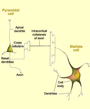
- Pyramidal cells: the principal output neurons of the cortex. They are rather unusual looking: picture a cute tepee shaped cell body with feelers (or “dendrites”) coming out of the top or apex (apical dendrites that extend towards the upper layer of the cortex) and the bottom corners or base (basilar dendrites that extend horizontally from the soma aka the cell body). The largest type of pyramidal cells are called Betz cells.
- Stellate neurons aka granular cells: highly dendritic, multipolar interneurons. These cells are typically small and act as the go between between efferent and afferent neurons.
- Cells of Martinotti: these cells are shaped like polygons. They form synapses with pyramidal cells.
- Fusiform cells: rod-shaped cells oriented perpendicularly from the surface of the cortex. They play a similar role to pyramidal cells.
- Horizontal cells of Cajal: these cells are small and rod-shaped. They are found in the most superficial layer of the cortex where they are oriented parallel to the surface. These cells disappear after birth.
When you recognize someone at a family gathering, come up with a witty comeback or decide to pass on another helping of dessert, remember all the neurons behind the scenes helping you to be you!