Most of us will remember from high school biology class that kidneys comprise part of the excretory system and function in toxin removal, maintaining electrolyte homeostasis and regulating the body’s acid-base balance. Beyond this, proper kidney function is also critical for the secretion of several important hormones such as erythropoietin and renin, which regulate red blood cell production and arterial blood pressure, respectively. Given the complex roles of the kidney, it’s no surprise that its structure is just as complex with many different parts and cell types working together to carry out its functions.
Kidney Structure Recap
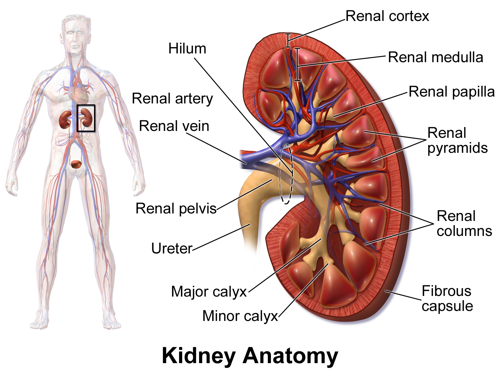
The kidney is organized into the outer renal cortex and the inner renal medulla (Figure 1). Spanning the cortex and medulla are approximately 2 million nephrons. These are complex epithelial tubules that constitute the kidney’s basic structural and functional units. Each nephron is subdivided into a filtration unit called the glomerulus, where filtration occurs, and a segmented tubular resorption compartment (Figure 2). The Bowman’s capsule is a watertight cup-shaped enclosure comprised of parietal epithelial cells, where primary urinary filtrate collects and is emptied through a network of epithelial tubules that starts at the proximal tubules, followed by the loop of Henle, the distal tubules, and finally a collecting duct.
  |
  |
| Figure 1: Kidney position and gross structure | Figure 2: Schematic representation of a nephron |
Nephrons filter approximately 20% of the blood volume that enters the kidneys, removing the waste products from amino acid and protein metabolism, while reabsorbing water and nutrients to maintain homeostasis. In this post, we take a look at just two of the cell types that make up the nephron – the glomerulus podocytes and the proximal tubules.
Glomerulus Podocytes
Structure and Function
Podocytes are one of four cell types within the glomerulus of the nephron, and are a major component of the ultrafiltration machinery. They are terminally differentiated and highly specialized perivascular cells composed of a cell body and elaborate projections known as major processes, that intertwine with each other to form foot processes, which wrap around and encase the glomerular capillaries and neighbor the cells of the Bowman’s capsule.
A well functioning kidney filters metabolic byproducts into the urine while blocking the passage of larger essential molecules such as albumin. This selective filtration process occurs across the glomerular capillary wall, and podocytes play an active role in this process. When foot processes from adjacent podocytes intertwine with each other, they form a network of narrow and somewhat uniform gaps at the glomerular basement membrane (GBM). The porous capillaries of the glomerulus thus retain blood cells and permit the passage of small solutes, while the overlying GBM is less permeable to macromolecules, in particular to albumin. The cytoskeletal dynamics, structural plasticity of and signaling between podocytes is essential to their role in glomerular filtration and proper kidney function.
Proximal Tubules
Structure and Function
The proximal tubule lies in the kidney’s cortex, and is the part of the nephron that begins at the urinary end of the glomerulus and ends at the loop of Henle. The tubule is seen as three segments; S1, S2, and S3, where S1 and S2 make up the proximal convoluted tubule that comprises complex cells, while the cells that make up S3 constitute the proximal straight tubule and are less complex.
The proximal tubule is distinguishable by its luminal brush border, and its roles are diverse, spanning regulatory, endocrine and immune functions. The proximal tubule is the major site of reabsorption in the nephron, where the largest fraction of glomerular filtrate is reabsorbed, including glucose, amino acids and calcium, as well as other divalent ions. The proximal tubule can also reabsorb small proteins and peptides via endocytosis. The ability to reabsorb is facilitated largely by the tightly packed microvilli that adorn the luminal surface of the epithelial cells of the proximal tubules. The cytoplasm of these epithelial cells is packed with mitochondria, primarily in the basal region within the infoldings of the basal plasma membrane. The many mitochondria provide energy for the active transport of sodium ions out of the cells, thus creating a concentration gradient that permits more sodium ions to enter the cell from the luminal side, while water passively follows the sodium out of the cell along the same concentration gradient.
Kidney Disease
Podocytes
Inherited or acquired impairment or damage of podocytes may lead to foot process effacement which is a morphological hallmark of proteinuric kidney diseases, for example, diabetes and minimal change disease. During effacement, podocytes lose their structure and spread out, resulting in reduced filtration barrier function. Effacement is thought to the result of a breakdown in the actin cytoskeleton of the foot processes, thus hampering the ability of the podocytes to reorganize themselves according to fluctuations in filtration needs. The implication of podocytes in kidney disease has been recently reviewed elsewhere (1).
Proximal Tubules
Cells of the proximal tubules are also implicated in many types of kidney disease. Recent years have witnessed a shift in the focus from the glomerulus and the kidney interstitium to the proximal tubules as a major component of kidney disease. High rates of oxygen consumption and their inherent antioxidant defense activity leave the cells of the proximal tubule particularly susceptible to injury. Recent research has revealed significant involvement of the proximal tubule in the progression from acute to chronic kidney disease, and most cases of renal cell carcinoma (RCC)- the most common type of kidney cancer – arise from the proximal tubules. For a recent review on the proximal tubule and kidney disease, check out Chevalier (2016) (2).
(UPDATE: After this blog post went LIVE, human iPSC-derived proximal tubules and podocyte 3D spheroids became available (Tempo Bioscience, San Francisco, California). Check out the announcement here!)
References
1. P. Garg, A Review of Podocyte Biology. Am J Nephrol 47 Suppl 1, 3-13 (2018).
2. R. L. Chevalier, The proximal tubule is the primary target of injury and progression of kidney disease: role of the glomerulotubular junction. Am J Physiol Renal Physiol 311, F145-161 (2016).
Figure Credits
Figure 1: Kidney Anatomy. Blausen.com staff (2014). "Medical gallery of Blausen Medical 2014". WikiJournal of Medicine 1 (2). DOI:10.15347/wjm/2014.010. ISSN 2002-4436. [CC BY 3.0 (https://creativecommons.org/licenses/by/3.0)]
Figure 2: Physiology of Nephron. Madhero88 [CC BY 3.0 (https://creativecommons.org/licenses/by/3.0)]
Article by Karen O’Hanlon Cohrt PhD. Contact Karen at karen@tempobioscience.com.
Karen O’Hanlon Cohrt is a Science Writer with a PhD in biotechnology from Maynooth University, Ireland (2011). After her PhD, Karen moved to Denmark and held postdoctoral positions in mycology and later in human cell cycle regulation, before moving to the world of drug discovery. Her broad research background provides the technical know-how to support scientists in diverse areas, and this in combination with her passion for writing helps her to keep abreast of exciting research developments as they unfold. Follow Karen on Twitter @KarenOHCohrt. Karen has been a science writer since 2014; you can find her other work on her portfolio.