by Karen O'Hanlon Cohrt | Mar 22, 2026 | Disease Models, Trends
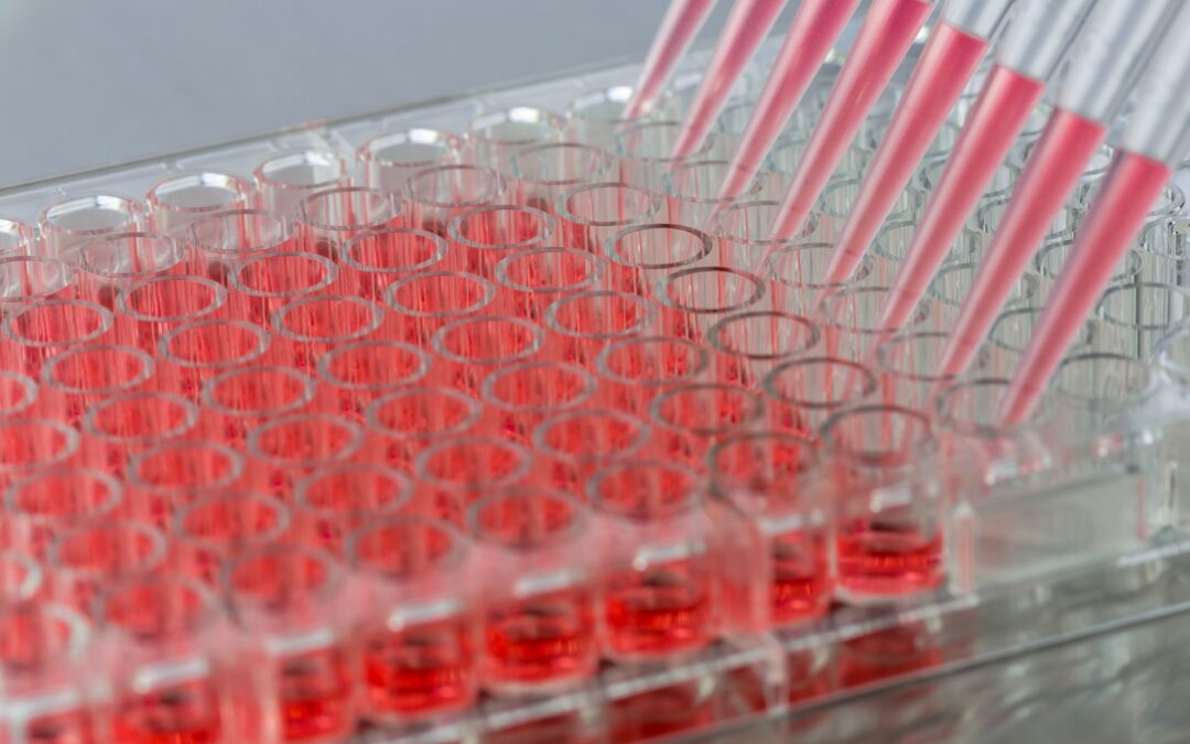
It is no secret that cell-based models are the backbone of modern drug discovery. For decades, researchers have used animal-based models including cells and whole animals to investigate mechanisms of disease, screen compounds, and support preclinical research. Over...
by Karen O'Hanlon Cohrt | Jan 6, 2026 | Cell Culture Techniques, Disease Models
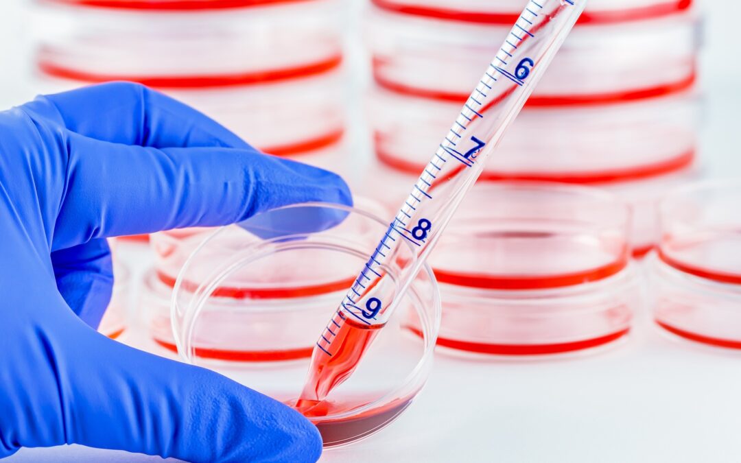
What is the difference between ECM-based and ECM-free systems in 2D and 3D cell culture? When are ECM-derived cues required for tissue organization and differentiation in 3D models? How do researchers choose between ECM-based and ECM-free culture formats for their...
by Karen O'Hanlon Cohrt | Nov 6, 2025 | Cell Culture Techniques, Disease Models
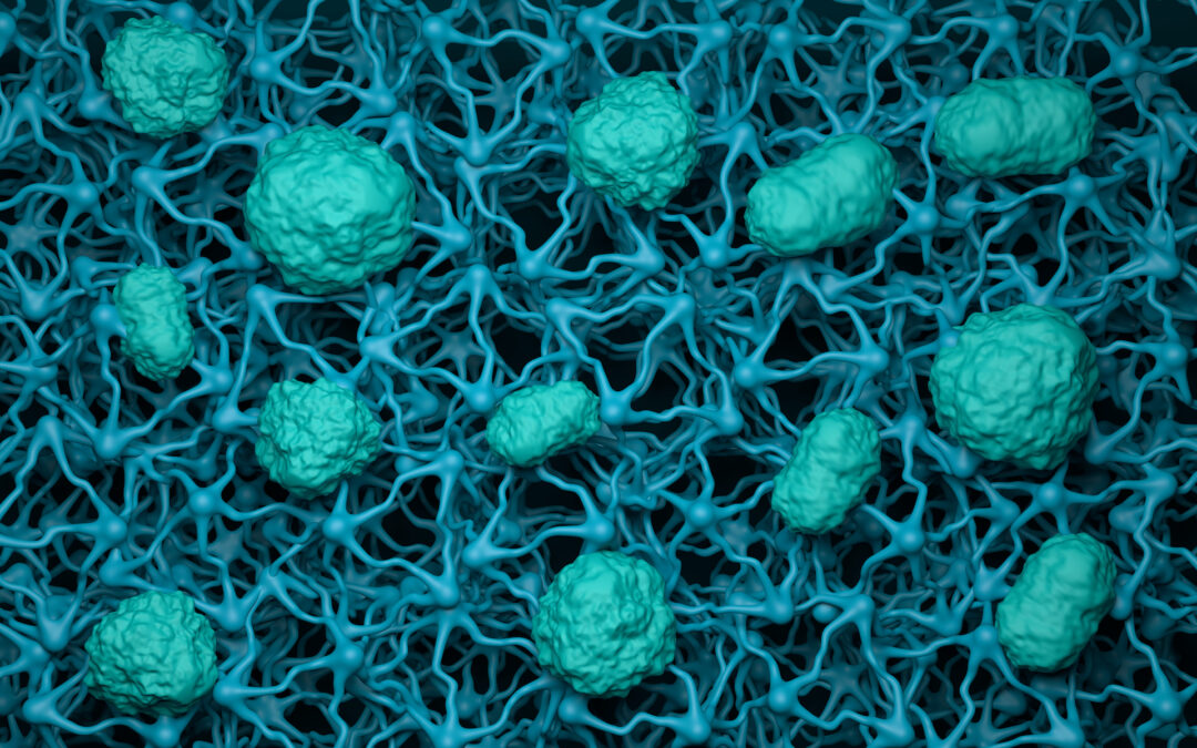
How do Transwell assays help model biological barriers in the lab? What does TEER tell us about barrier integrity and tight-junction strength? How can these assays advance our understanding of drug transport and disease? Biological barriers are critical for...
by Karen O'Hanlon Cohrt | Oct 6, 2025 | Disease Models, Trends
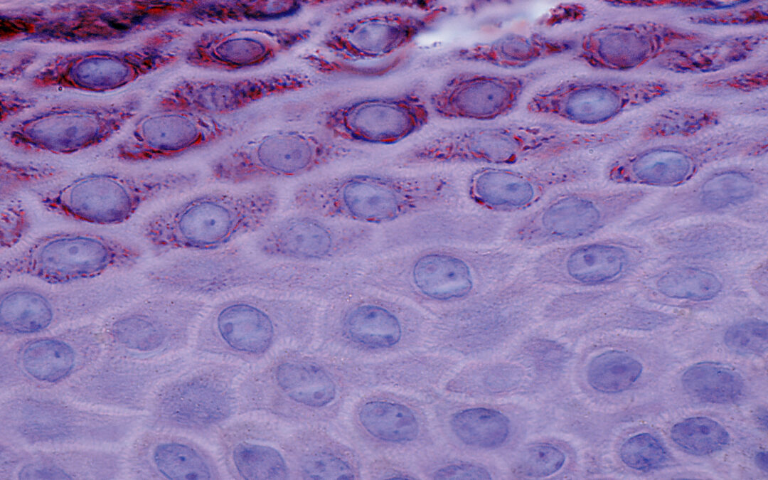
In a previous article we introduced keratinocytes and looked at their biological functions and subtypes. Here, we explore some of the main reasons researchers study keratinocytes and the various approaches used. We focus on 2D assay formats, outlining their advantages...

by Karen O'Hanlon Cohrt | Jul 13, 2025 | Cell Culture Techniques, Disease Models
In our last article, we compared 2D organ-on-a-chip devices and 3D organoids with respect to their use in drug discovery, highlighting their importance in modeling diseases and evaluating efficacy and safety during drug discovery and development . We also presented...